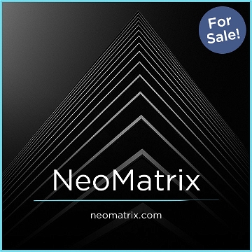
|
|
|
|
This content requires the Macromedia Flash Player.
Get Flash
|
 |
|
|
Breast Cancer Development
To appreciate the concept of screening for benign breast disease, and the
analogy to the Pap test, it is important to understand the disease and it’s
course of development. Virtually all breast cancer originates in the epithelial
cells that line the interior of the milk ducts in the breasts. Like cervical
cancer, breast cancer progresses through identifiable stages of development.
Like cervical cancer, breast cancer typically grows slowly, taking, on average,
8 years before it can be detected by mammography, or up to 10 years before the
lesion is palpable. The inability of current breast screening methods to detect
cellular changes at an early stage of development, as the Pap test does for
cervical cancer, is one reason for the lack of significant reduction in the
death rate of breast cancer.
A major difference between the cervical Pap test and NAF cytology is that most
asymptomatic women do not secrete NAF, or secrete acellular samples. These are
normal states and confer the lowest risk of developing breast cancer. With the
cervical Pap test, it may be reported that the sample is inadequate for
analysis if very few cells are present. In NAF cytology this is reported as an
acellular sample, which confers the lowest relative risk among patients who are
secretors. Multiple studies have confirmed that women who do not produce a NAF
sample are at a significantly reduced risk of developing breast cancer,
compared to women with abnormal cytology.
Clinical Utility of Screening for Benign Breast Disease
With the understanding that almost all breast cancers begin in the milk
ducts, Nipple Aspirate Fluid (NAF) examination allows the determination of
benign breast disease, and the attendant increase in relative risk, years
before an abnormality becomes visible by imaging or becomes a palpable lesion.
The study of NAF to identify high risk women is not new. In 1958, Dr. George
Papanicolaou et al described obtaining fluid from the breast milk ducts by
suction to analyze cell samples. Just like his procedure for detecting normal
versus abnormal cells in the cervix (the "Pap test"), this technique
demonstrated the ability to find abnormal cells in nipple aspirate fluid (NAF)
from within the breast duct. Dr. Papanicolaou concluded that "cytology of
breast secretions was valuable in differential diagnosis of mammary diseases
and carcinoma" and that "a cytological diagnosis of malignancy was highly
reliable."
Multiple clinical studies involving over 30,000 patients, followed for up to 25 years,
all reach the same
conclusion: benign breast disease, specifically atypia, confers a significantly
higher risk of breast cancer. On average, these clinical studies demonstrate
the presence of atypia to mean a woman has a greater than 4X (i.e., 400%)
relative risk of developing breast cancer than women who do not produce fluid.
The findings of Wrensch, Petrakis et al and the clinical significance of
atypical hyperplasia have been validated through independent studies of samples
collected from NAF, fine needle aspiration biopsy, or surgical excision.
An official statement from the American Society of Breast Surgeons reads in
part "A variety of techniques exist to obtain cells for examination, including
open surgical biopsy, fine needle aspiration (FNA), nipple aspirate fluid
(NAF), and ductal lavage. In current studies of surgical biopsy, FNA and NAF
report a 5-fold increase in the relative risk of developing breast cancer when
cellular atypia is found. Having atypia coupled with a first degree family
history of breast cancer confers an 11 to 22-fold increase in relative risk of
developing breast cancer."
Conclusion. We have known for decades that atypia is an important breast cancer
risk factor, but have lacked a practical way to apply this knowledge to the
general asymptomatic population. The primary care setting is the point-of-care
where screening must occur, but the breast specialist must manage the high-risk
patients. With the advent of a noninvasive, office-based
method of collecting NAF, we have the opportunity to enter more high risk women
into enhanced surveillance programs earlier, and help them avoid a
life-threatening battle with breast cancer.
Click to view
Screening for Benign Breast Disease
Abstracts Summary
Key Articles:
- West J.G., Hollingsworth A.Screening for breast cancer risk in the obstetric/gynecological setting: a breast surgeon's perspective Exp Rev. Obstet. Gynecol. 2008, 3(1), 59-63
- Medco Forum HALO Breast Pap Test By NeoMatrix Medco Forum 2008; 15(17), 1-2
- Wrensch MR, Petrakis NL, King EB, et al. Breast cancer risk in women with abnormal
cytology in nipple aspirates of breast fluid. American Journal of Epidemiology 1992;135(2):130-141
- Tice JA, Miike R, Adduci K, et al. Nipple Aspirate Fluid Cytology and the Gail Model
for Breast Cancer Risk Assessment in a Screening Population. Cancer Epidemiol
Biomarkers Prev 2005;14(2):324-328.
- Hartmann LC, Sellers TA, Frost MA, et al. Benign Breast Disease and the Risk of
Breast Cancer, New England Journal of Medicine 2005; Volume 353:229-237 Number 3
- Hollingsworth A, Singletary S, Morrow M, et al. Current comprehensive assessment and
management of women at increased risk for breast cancer, The American Journal of
Surgery 187 (2004) 349-362
- Proctor K, Rowe L, Bentz J. Cytologic features of nipple aspirate fluid using an
automated non-invasive collection device: a prospective observational study, BMC
Women's Health 2005, 5:10
- Lee WY. Cytology of abnormal nipple discharge: a cyto-histological correlation,
Cytopathology 2003, 14, 19-26
Back to top
|
|
|
|
|
|